| Tick Patrol HLFS Ursprung 2006 |
|
| Home | |
| Abstract | |
| Motivation | |
| Ticks | |
| The Hunt | |
| Laboratory Analysis | |
| Assignment | |
| Video | |
| Bibliography | |
| Team | |
| Imprint | |
![]()
 |
|
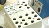
| The enzymes need time and proper temperature to take effect. |
 |
|
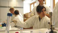
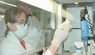
| Women in engineering—having lots of fun. |
 |
|
| extreme concentration. |
 |
|
| Difficult, but not impossible. |
 |
|
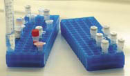
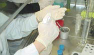
At over 20 reagents organization is absolutely essential. |
 |
|
| Careful disinfection before and after each session. |
 |
|
| Used material was autoclaved. |
 |
|
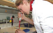
| Working with micro-eye-droppers is a big challenge. |
Laboratory Analysis
Detection of Lyme disease and encephalitis in ticks.
The pathogens, which causes Lyme disease are bacteria, so called borrelia (spiral-shaped bacteria). The genetic material of borrelia is DNA.
The cause of TBE is a virus. The genetic material of the TBE-virus is RNA.
It is now possible to detect both TBE and Lyme disease pathogens with a DNA test done directly on the tick.
This test provides timely results in contrast to the serological laboratory tests, for example the ELISA-TEST, which detects antibodies in the blood of the patient only after a few weeks. In very serious cases the treatment can be applied immediatly.
In preparation for work done in the school laboratory, the captured ticks are stored for several weeks in 70% alcohol at a temperature of -70°C. After this period of time we can be sure that the alcohol has killed all disease-causing microorganisms, so that there is no danger of infection. Storing the ticks at such an extremely low temperatur assures that the genetic material of the pathogens won’t be broken down by enzymes. A professional laboratory with a staff trained to handle pathogens in a safe manner would begin right away to work with the ticks.
We start by detecting borrelia and TBE viruses in the ticks with the help of the DNA\RNA analytics. This means that a test is done to see if the genetic material of the given pathogen can be detected in the sample, (in this case being the tick).
This means that in order to potentially detect these pathogens the borrelia-DNA as well as the TBE-RNA must be isolated. The normal procedure would be to buy a kit that already contains a detailed protocol for isolating either DNA or RNA. Since in this special case every tick was to be tested not only for borrelia, but also for TBE viruses, an isolation protocol for both nucleic acids (DNA+ RNA) had to be found. We finally succeeded to find an appropriate method: “MasterPure Complete DNA & RNA” from the US company Epicentre, which proved to be suited to our purposes.
The first step in extracting the genetic material of the pathogens is to mechanically destroy the chitin shell of the ticks either by pulverizing it in liquid nitrogen or with the use of an Ultraturrax, a special kind of mini turbo-powered hand-held blender with over 20,000 rotations per minute. The cells are then lysed with special enzymes. Fats, proteins, cell-organelle and salts that would otherwise interfere are removed in several consecutive purification processes. After the isolation of the genetic material is complete, potential borrelia-DNA and TBE-virus-RNA is identified with the help of the so called PCR method.
The Polymerase Chain Reaction (PCR) plays an important part in the current routine diagnostics for the purpose of detecting bacteria or viruses and it allows the specific amplification of the least possible amounts of DNA of the pathogen in question.
Borrelia can be relatively easily detected using the PCR method (ist relativ einfach mit einem PCR –Schritt möglich), because the genetic material in this case consists of DNA. A suitable sequence for the amplification of a borrelia-gene was chosen from various sources of scientific writings.
In order to detect TBE, the virus-RNA must first be transcribed into DNA (reverse transcription), so that they can subsequently be amplified in a “nested PCR-process”. In the „nested-PCR” two PCR-rounds are carried out, one after the other, so that even the least possible amounts of the desired information can be traced. A somewhat smaller fragment of the first batch is duplicated in the second PCR-round. The „nested PCR” inhances the sensitivity und specificity of the detection process.
This process is termed RT-PCR (Reverse Transcriptase - Polymerase-Chain Reaction): First the Reverse Transkriptase (RT) is employed to transcribe the RNA in cDNA (c stands for complementary). This cDNA can subsequently be used in a PCR to amplify the sequences in question.
The products of the PCR are then analysed on an agarose gel, through which the DNA-fragments can be sorted and evaluated according to size.
The subsequent interpretation of the results is relatively easy. A negative PCR-result indicates that genetic material from the pathogens cannot be found in the tick. Risk of infection can thereby be almost completely ruled out. A positive result on the other hand proves the presence of borrelia bacteria or TBE-viruses resp. and the risk of a consequent transmission of the disease-causing microorganism in the case of a tick bite.
A positive control was carried out in order to assure that no mistakes were made in the process of the diagnostic analysis. RNA from the tick’s mitochondria from each sample was also transcribed into cDNA (reverse transcription) and was then amplified with a PCR. In order to assure reliable results all control reactions had to provide a visible PCR-product, whereas in the case of the borrelia and TBE-PCR only infected ticks would have to provide a signal when analysed on the agarose gel. In the case of a mistake in the laboratory procedure no positive control would be detectable. This would indicate that the procedure would have to be repeated.
The entire process of detection was carefully verified using original borrelia-DNA, TBE-RNA and TBE c-DNA. Dr. Stephan Aberle, from the Clinical Institute for Virology at the Medical University of Vienna provided us with reference samples of TBE-virus genetic material. Dr. Arnold Bito, from the Cell Biology department at the University of Salzburg provided us with the reference samples of genetic material from borrelia.
1. A Summary of the Test Procedure:
· Isolation of the ticks’ DNA and RNA
· Selective detection of borrelia DNA with the help of the Polymerase-Chain Reaction (PCR)
· Reverse Transcription of the isolated RNA into cDNA (complementary DNA)
· Selective identification of FSME-viral cDNA with the help of Polymerase-Chain Reaction (PCR)
· Identificaiton of tick-cDNA as positive control
· DNA-Gel-Electrophoresis for visualization of the PCR product and evaluation of the results.
2. The Experimental Procedure – The Laboratory Protocol
2.1 DNA and RNA Isolation
(MasterPure Complete DNA and RNA Purification Kit, Fa. Epicentre)
· Disrupt the chitin shell of the ticks using either liquid nitrogen or the Ultraturrax for mechanical disruption
· Add 300µl Tissue and Cell Lysis Solution
· Add 1µl Proteinase K
· Vortex
· Incubate 15min at 65°C; vortex every 5 minutes (The cells will lyse)
· Store on ice for 3min
· Add 160µl MPC Protein Precipitation Solution (Proteins are destroyed and precipitate)
· Vortex for 10sec
· Centrifuge for10min (to get rid of the proteins)
· Transfer the supernatant (contains the nucleic acids) into a new test tube
· Add 500µl ice cold Isopropanol, tilt the test tube 30-40x to mix
· Centrifuge 10min (the nucleic acids are now in the pellet!)
· CAREFULLY drain off the alcohol
· Wash the pellet (DNA and RNA!!!) with 150µl 75% EtOH
· Drain the alcohol completely, briefly dry the pellet
· Dissolve the DNA and RNA in 35µl of water
4.2.2 Amplification of the Borrelia DNA using PCR
(Primer: Priem S, Rittig MG, Kamradt T, Burmester GR, Krause A.)An optimized PCR leads to rapid and highly sensitive detection of Borrelia burgdorferi in patients with Lyme borreliosis.
J Clin Microbiol. 1997, 35(3):685-90.)
Protocol for the Amplification of the Borrelian Gene p66:
Product consists of @ 400 bp
ddH2O 36.8 µl
PCR buffer 10x 5 µl
dNTPs 1 µl 10 mM
Primer 1 1 µl 10 pmol/µl
cgaagatactaaatctgt
Primer 2 1 µl 10 pmol/µl
gatcaaatatttcagctt
Sample (DNA) 1 µl MgCl2
4 µl 25 mM Taq polymerase 0.2 µl 2U/µl
PCR-Conditions:
initial 3 min 94°C,
Then 35 cycles:
94 °C.......30 sec
45 °C.......30 sec
72 °C.......30 sec
2.3 Amplification of TBE-RNA and positive control through RT-PCR
(RevertAid First Strand cDNA Synthesis Kit, Fa. Fermentas)
The first step was to transcribe the RNA through reverse transcription into cDNA. An extremely high amount of randomly generated primers garauntees that all of the available RNA (TBE-virus specific as well as tick-inherent) is transcribed for the positive control.
5 µl sample (RNA)
1 µl random hexamer primer
6 µl DEPC-treated water (contains no RNAsen!)
· Vortex briefly
· Incubate 5min 70°C, store briefly on ice
4 µl reaction buffer
1 µl Ribonuclease Inhibitor
2 µl dNTPs
· Vortex briefly
· 5min room temperature
· Add 1 µl Reverse Transcriptase
· 10min room temperature
· 60min 42°C
· Stop the reaction by heating 10min 70°C
In the next step an ordinary PCR follows, in which the newly created cDNA is used as a template. In this part the desired cDNA sequence is selectively amplified using the respective primers for the first time. Erst bei diesem Schritt wird selektiv mit passenden Primern die gesuchte cDNA Sequenz amplifiziert
The following takes place:
a.) TBE-Virus RNA (res. cDNA) and
b.) mitochondrial RNA (res. cDNA) of the ticks (serving as a positive control) are amplified.
a) Amplification of TBE-cDNA
(Primer from Dr. Stephan Aberle, Clinical Institute for Virology, Medical University Vienna)
In order to increase the specifity and sensitivity of the TBE- identification, a so-called nested-PCR is employed. The first PCR product serves as a template for the second round. The new primers are located further inside than the first primers, so that only the specific DNA segments of the first round are amplified. This increases the sensitivity and efficiency so greatly, that the technicians must work very carefully and must assure that no contamination takes place, since any sort of impurity could have an undesirable effect.
In the nested-PCR the first PCR product, after it has been successfully amplified for the first time, is used as a template for a second PCR.
1. PCR TBE:
10 µl cDNA (from 2.3)
1 µl primer1 (5´-gaggctgaacaactgcacga-3´)
1 µl primer2 (5´-gaacacgtccattcctgatct-3´)
1.5 µl dNTPs (10mM)
0.2 µl Taq Polymerase
5 µl buffer
4 µl MgCl2
27.3 µl H2O
25 cycles:
94 °C.......30 sec
40 °C.......30 sec results in a product of 357 bp
68 °C.......30 sec
2. PCR TBE:
2 µl of the first PCR
1 µl primer3 (5´-acggaacgtgacaaggctag-3´)
1 µl primer4 (5´-gcttgttaccatctttggag-3´)
1.5 µl dNTPs
0.2 µl Taq Polymerase
5 µl buffer
4 µl MgCl2
35.3 µl H2O
30 cycles:
94 °C.......30 sec
50 °C.......30 sec results in a product of 252 bp
72 °C.......30 sec
b) The Amplification of mitochondrial ticks’ cDNA as Positive Control.
(Primer according to. Info. from Dr. Judith Kießling, Institute for Medical Microbiologie, Infectious- und epidemic medicine, Veterinary Faculty, Ludwig-Maximilians-Universität München)
To assure that the isolation of the RNA as well as the reverse transcription were succesfully carried out, a certain segment of the mitochondrial tick-RNA and the respective cDNA is duplicated.:
PCR-mix:
2 µl cDNA (from 2.3)
1 µl dNTPs
1 µl primer 1
F-16S: aaaaaaatactctagggataacagcgtaa
1 µl primer 2
R-16S: accaaaaaagaatcctaatccaaca
5 µl buffer
4 µl MgCl2
0.2 µl Taq Polymerase
35,8 µl H2O
35 cycles:
94 °C.......30 sec
56 °C.......30 sec results in a product of 700 bp
72 °C.......30 sec
2.4 Visualization of all PCR-Products by Agarose Gel Electrophoresis
In order firstly to make the PCR-products visible and secondly to determine their size, an Agarose-gel-elektrophoresis is carried out.
DNA-gel:
1 % Agarose gel in 0,5x Tris-Borat-buffer pH 8,6 (TBE)
+ 8 µl dye (Gelstar)
DNA-dye (Gelstar): stained reagent that forms adducts on the DNA is induced to florescence.
Can be caused to glow when exposed to Ultraviolet light (UV), thereby making the individual DNA fragments visible in the gel.
Preparation of the sample for the Elektrophoresis:
50 µl sample (PCR)
10 µl sample buffer (BPB)
Sample buffer: contains a dye (= bromphenol blue), which makes it possible to follow the migration of the DNA in the gel and glycerin to “weigh down” the DNA, which subsequently sinks into the gel-pockets.
Pipet 25 µl of the sample in the gel-slots.
Duration of seperation at least 45 minutes at 100 mA
The DNA migrates from the negative to the positive pole; The migration of the DNA in the gel can be traced based on observations of the dye front. After approximately 45 minutes the gel is removed from the dye gel chamber and placed on the illuminator. The gel is observed and photographed on the blue-light illuminator. The stained DNA-bands show a greenish-yellow glow.
The marker provides information about the size of the bands.
This technique causes the longer fragments of DNA, while migrating through an electric field, to stay longer in the Agarose-network than the smaller fragments; thus allowing for the sorting of the particles according to size.